/filters:quality(90)/prod01/production-cdn-pxl/media/umassdartmouth/features/images/guo-students.jpg)
Iron is the most abundant, nutritionally essential metal found in the human body. It plays an important role in processes such as oxygen delivery, electron transport, enzymatic reactions, and DNA synthesis and repair.
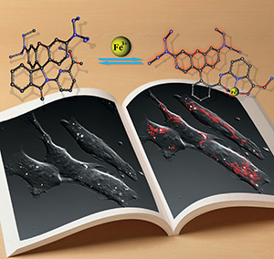
However, iron can also catalyze the production of free radicals, which attack living cells and are linked to several diseases. Both iron deficiency and iron overload are related to various health problems—yet effective tools for monitoring iron cells in biological systems have remained a challenge.
Dr. Maolin Guo, professor of chemistry and biochemistry, has recently developed a new method for monitoring iron in living human cells via imaging probes, which could lead to better detection and treatment of diseases such as cancer, neurodegenerative diseases, hemochromatosis, and cardiovascular diseases. The probes generate a fluorescent response within the cells to let scientists monitor iron levels more effectively.
New imaging probes detect iron pools in living cells
Precisely monitoring iron ions (Fe2+ and Fe3+) in the human body and other biological systems is important in understanding the detailed biological functions of iron and its trafficking pathways.
Dr. Guo's lab has developed chemically-based probes that produce a fluorescent signal, based on the interactions between the probes and the iron ions in the living cells. The detection and mechanism of the interaction were verified by the magnetic properties of the probes using an E-Scan Electron Paramagnetic Resonance (EPR).
"We are quite excited about the new imaging probes," Dr. Guo said. "The probes we developed, combined with the new tools we have utilized, enable us to precisely monitor iron ions at subcellular resolution in live cells."
Making the connection between the classroom and the lab
Making the connection between the lab and the classroom is an important aspect of Dr. Guo's work.
"I teach undergraduate and graduate biochemistry, modern bioanalytical techniques, and advanced cell and molecular biology courses in which I introduce my research to students," he said.
"Last semester, I gave a seminar in our chemistry seminar course where I presented my research to the undergrad and graduate students in our department. Students usually do their graduate or undergraduate research projects with me in my lab. I also have undergraduate students supported by the Office of Undergraduate Research and summer interns."
Dr. Guo noted that research opportunities are open to both undergraduate and graduate students not only in chemistry, but also in biology, medical laboratory science, and bioengineering.
The interface of chemistry, biology, and medicine
The National Science Foundation has funded Dr. Guo's work through two grants totaling $739,000. His lab received $400,000 to develop the iron imaging probes and a $339,000 grant to support the acquisition of a 400 MHz digital Nuclear Magnetic Resonance (NMR) spectrometer and an E-Scan Electron Paramagnetic Resonance (EPR) spectrometer which played critical roles in this research.
 Dr. Guo and his fellow researchers from UMass Dartmouth's Biomedical Engineering and Biotechnology PhD program, the UMass Cranberry Health Research Center, and UMass Amherst recently published an article on their work in Dalton Transactions, a front-line journal for inorganic, organometallic, and bioinorganic chemistry. Dr. Guo was also recognized this year by UMass Dartmouth's Faculty Federation as Scholar of the Year.
Dr. Guo's research is focused on the interface of chemistry, biology, and medicine. This exciting field is the foundation of modern biomedical sciences which provide molecular basis for various biomedical processes.
The research team uses a multi-disciplinary approach including molecular biology, protein chemistry, synthetic and electro-chemistry, spectroscopy (NMR, UV-vis, MS, fluorescent, etc), microscopy and X-ray crystallography to investigate the molecular basis of certain genes, proteins, metal ions, reactive oxygen species (ROS) and signal transduction pathways related to cancer, aging, infectious, cardiovascular diseases and neurodegenerative disorders.
The goal is to understand the molecular basis of these biochemical processes and to develop new strategies for diagnostics and therapeutics. Current research activities are focused on molecular imaging probes for iron and ROS detection in living systems as well as the health benefits of cranberry and related natural products.
More information
Department of Chemistry & Biochemistry
Biomedical Engineering & Biotechnology PhD Program
UMass Cranberry Health Research Center
In the news: Ribbon cutting for 400 MHz Nuclear Magnetic Resonance (NMR) facility